Iron Deficiency Anemia Peripheral Blood Film

Peripheral Blood Film With Changes Attributed To Iron Deficiency Anemia Download Scientific Diagram

Iron

The Peripheral Blood Film In Severe Iron Deficiency Anaemia The Cells Download Scientific Diagram

File Iron Deficiency Anemia Peripheral Blood Smear 4422704616 Jpg Wikimedia Commons
Iron Deficiency Anemia
Laboratory Diagnosis Of Iron Deficiency Anemia Ida
Different tests help your doctor diagnose iron deficiency anemia.
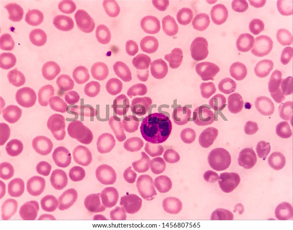
Iron deficiency anemia peripheral blood film. Peripheral smear to see if your red blood cells look smaller and paler than normal when viewed under a microscope. Here is another peripheral blood smear demonstrating changes with iron deficiency anemia. In sideroblastic anaemia there is increased bone marrow iron. Moderate iron deficiency.
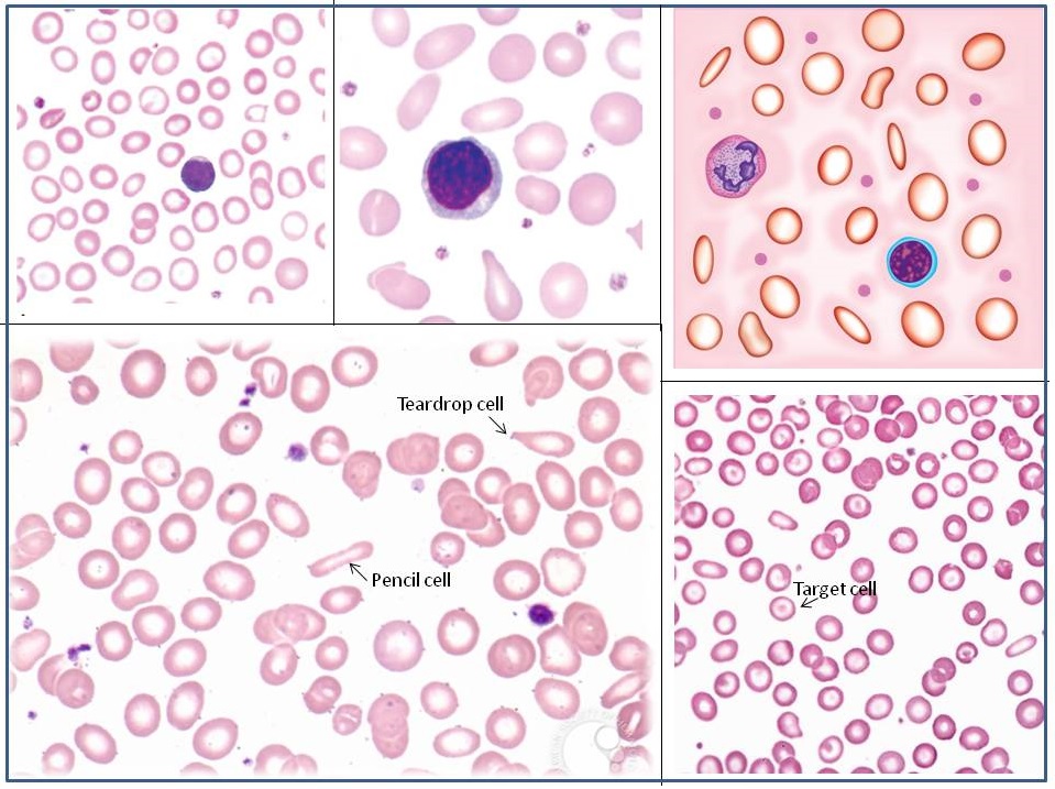
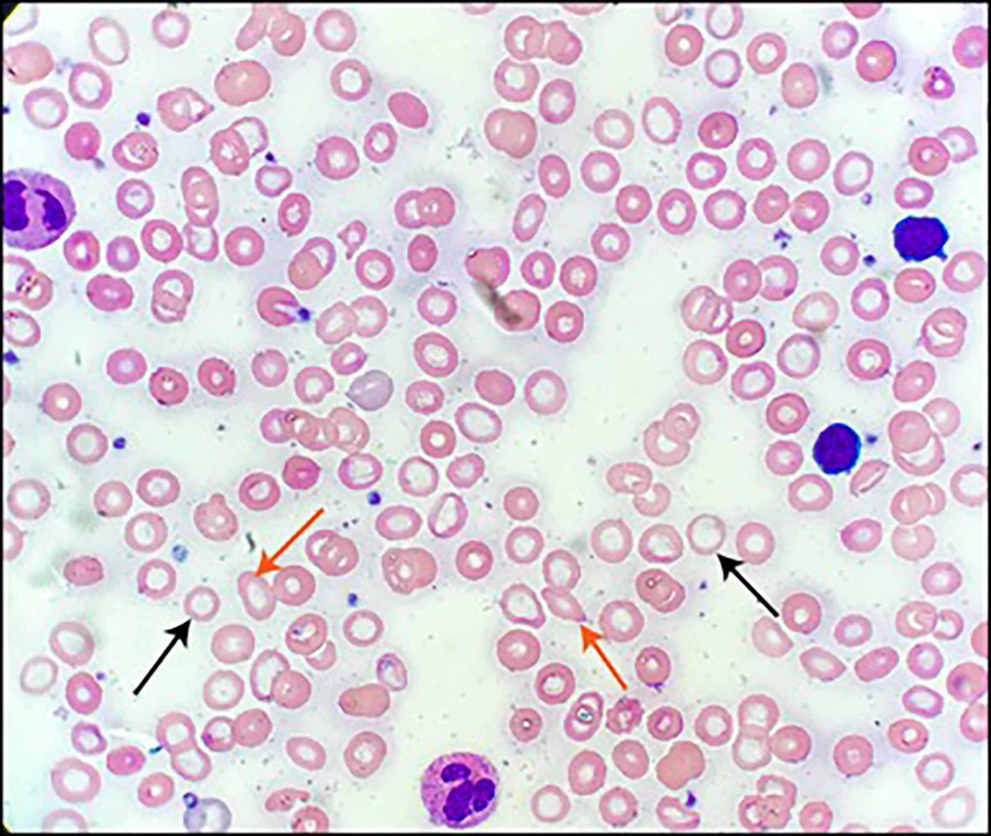
A peripheral blood smear looks at the size and shape of your red blood cells. Examination of the erythrocytes shows microcytic and hypochromic red blood cells in chronic iron. In iron deficiency anemia blood levels of iron will be low or less than 10 micromoles per liter mmol l. A high mcv with normal folate and b12 levels normal iron and a blood film showing anisocytosis and poikilocytosis suggests sideroblastic anaemia.
Sideroblasts are abnormal red cell precursors with iron loaded mitochondria forming a ring around the nucleus. Examination of the peripheral smear is an important part of the workup of patients with anemia. Except when morphologic changes are pronounced the diagnosis of iron deficiency anemia from examination of the peripheral blood film is difficult and not very reliable. Four of the participants examined the same slides twice in different random series and on the average contradicted 22 of their own interpretations.
With more severe iron deficiency anemia the peripheral blood smear may show hypochromic pencil shaped cells and occasionally small numbers of nucleated red blood cells. Significant hypochromia and microcytosis is seen as well as moderate variation in size and shape of the red cells. Normal lymphocyte for comparison purposes is seen at the edge of the smear. Note the increased zone of central pallor and the more irregular shapes of the rbc s.
Cureus Zinc Deficiency Induced Hypogeusia In A Patient With Refractory Iron Deficiency Anemia A Case Report
Iron Deficiency Anemia

Anemia Wikipedia

Iron Deficiency Anemia Hematology And Oncology Msd Manual Professional Edition

File Iron Deficiency Anemia Jpg Wikimedia Commons

Iron Deficiency Anemia Ask Hematologist Understand Hematology

Peripheral Blood Evaluation B12 Deficiency Anemia Et Cmml Cap Today

Irem A Kilic On Twitter Severe Iron Deficiency Anemia Showing Hypochromic Pale Microcytic Rbcs In Peripheral Blood Smear Hematology Pathology Hempath Photocredit Pambuccian Https T Co Eregs975rd
Peripheral Blood Film Iron Deficiency Anemia 1000 Oil Download Scientific Diagram

Blood Smear Iron Deficiency Uptodate

Microcytic Hypochromic Anemia Blood Smear And Cbc Reports Medical Laboratories

Irem A Kilic On Twitter Severe Iron Deficiency Anemia Showing Hypochromic Pale Microcytic Rbcs In Peripheral Blood Smear Hematology Pathology Hempath Photocredit Pambuccian Https T Co Eregs975rd